The Art of Anatomy - Wilhelm Röntgen's X-ray
The first scan of the human body was both a medical breakthrough and an early international media sensation
If you’ve ever benefitted from an X-Ray or an MRI or CT scan, you should thank one 19th century German scientist, and his accommodating wife.
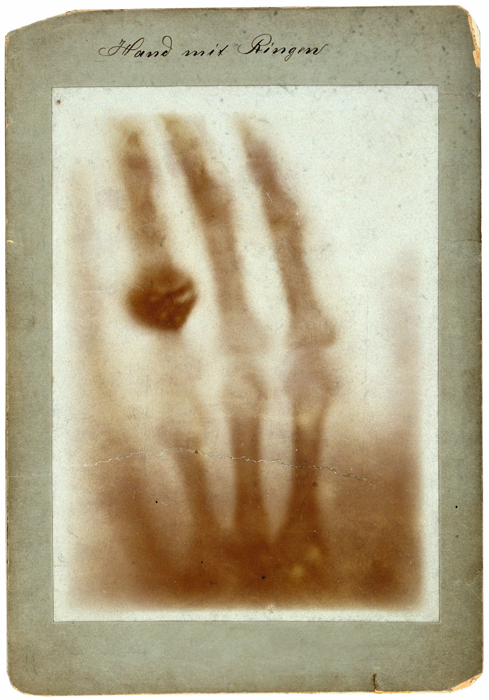
“The first X-ray photograph ever taken of the body, Hand with Rings, is the famous origin of all modern medical imaging,” explains our new book Anatomy: Exploring the Human Body. “Having discovered in November 1895 that substances opaque to visible light would transmit ‘a new kind of rays’, the German physicist Wilhelm Conrad Röntgen of the University of Würzburg worked hard for several weeks, then used those rays to view his wife Bertha’s hand with the rings symbolizing their marriage.”
Röntgen must have realised the scientific implications of his discovery. However, it is less likely that the physicist foresaw how his striking new image would also become an early international media sensation, demonstrating the ever-broadening reach of Victorian communications technology.
“On New Year’s Day 1896, he sent colleagues this and other photographs with offprints of his unillustrated article. A Viennese recipient told a physicist friend whose father edited a newspaper, which the next day reported what it called a ‘sensational discovery’: ‘Most amazing is the picture … of a human hand’, in which ‘rings appear to float freely …. The soft parts … are not visible.’ ‘Taken seriously by serious scholars’, the news was cabled around the world and the hand made the medical applications of the new technique clear.
“The print shown here had already been sent to Arthur Schuster in Manchester, England where his assistant translated Röntgen’s German into English for publication in Nature alongside a reproduction of this photograph and one of a flood of pictures of other hands. There would follow images of less accessible body parts, first using X-rays and later in the twentieth century also with technologies such as ultrasound, computerized tomography (CT), magnetic resonance imaging (MRI) and positron emission tomography (PET).”
Both in terms of modern medicine, and 19th century media reach, Bertha’s wedding ring in Wilhelm’s picture has truly encircled the globe.
To see how this image takes its place in the wider culture of visualizing the human body, get a copy of Anatomy here. The book is a visually compelling survey of more than 5,000 years of image making. Through 300 remarkable works, selected and curated by an international panel of anatomists, curators, academics, and specialists, it chronicles the intriguing visual history of human anatomy, showcasing its amazing complexity and our ongoing fascination with the systems and functions of our bodies. The 300 entries are arranged with juxtapositions of contrasting and complementary illustrations to allow for thought-provoking, lively, and stimulating reading. Buy your copy here.