The Art of Anatomy – Mark Fairchild
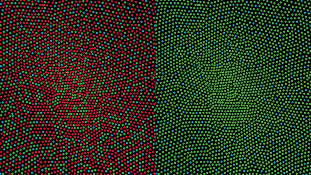
Our ability – and inability – to see colour is laid bare in this illustration of two very different human retinas
How do we see colour? As with phone screens, paint palettes and colour films, we build up a huge array of pigments from simpler blocks. “Each colour has a specific wavelength to which receptors or cones in the eye’s light-sensitive layer, the retina, are responsive,” explains the text in our new book Anatomy: Exploring the Human Body. “Three types of cones – for short (S, blue), medium (M, green) and long (L, red) wavelengths – enable normal trichromatic colour vision.”
However, a few individuals miss out on a key element within that range. In this illustration by Mark Fairchild, director of the Munsell Color Science Laboratory at the Rochester Institute of Technology, the retinal make-up of a healthy eye is set beside one missing a key attribute.
“The image on the left represents a mosaic of cone receptors at the fovea of a trichomatic eye,” explains the text. “There are around 150,000 receptors per square millimetre at the fovea, which is the retinal area with best colour vision. In the image on the right, the red (L) cones have been replaced with green (M) cones, signifying that the individual has a colour-vision deficiency known as red–green colour blindness.

“These individuals confuse bright reds and greens and cannot clearly distinguish colours containing red. Congenital red-green colour blindness affects about eight per cent of males and less than one per cent of females, but is passed from mother to son on the X chromosome.”
Can’t make out any major difference between those two pictures? Maybe it's time for that eye test!
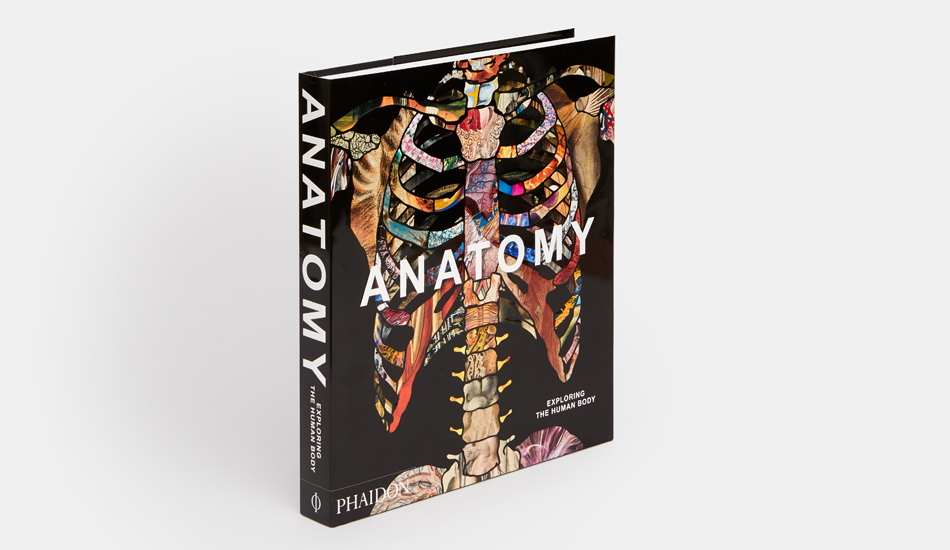
To see how this image takes its place in the wider culture of visualizing the human body, get a copy of Anatomy here. The book is a visually compelling survey of more than 5,000 years of image making. Through 300 remarkable works, selected and curated by an international panel of anatomists, curators, academics, and specialists, it chronicles the intriguing visual history of human anatomy, showcasing its amazing complexity and our ongoing fascination with the systems and functions of our bodies. The 300 entries are arranged with juxtapositions of contrasting and complementary illustrations that make for thought-provoking and lively reading. Buy your copy here.