The Art of Anatomy - The Anatomage Table
This touch-screen human cadaver enables students to get under the skin without reaching for the scalpel
Getting inside the human body has never been easier. Once, as Professor Thomas Schnalke explains in his introduction to Anatomy: Exploring the Human Body, Medieval medical students and practitioners, had to dissect and observe human cadavers in operating theatres, or even churches.
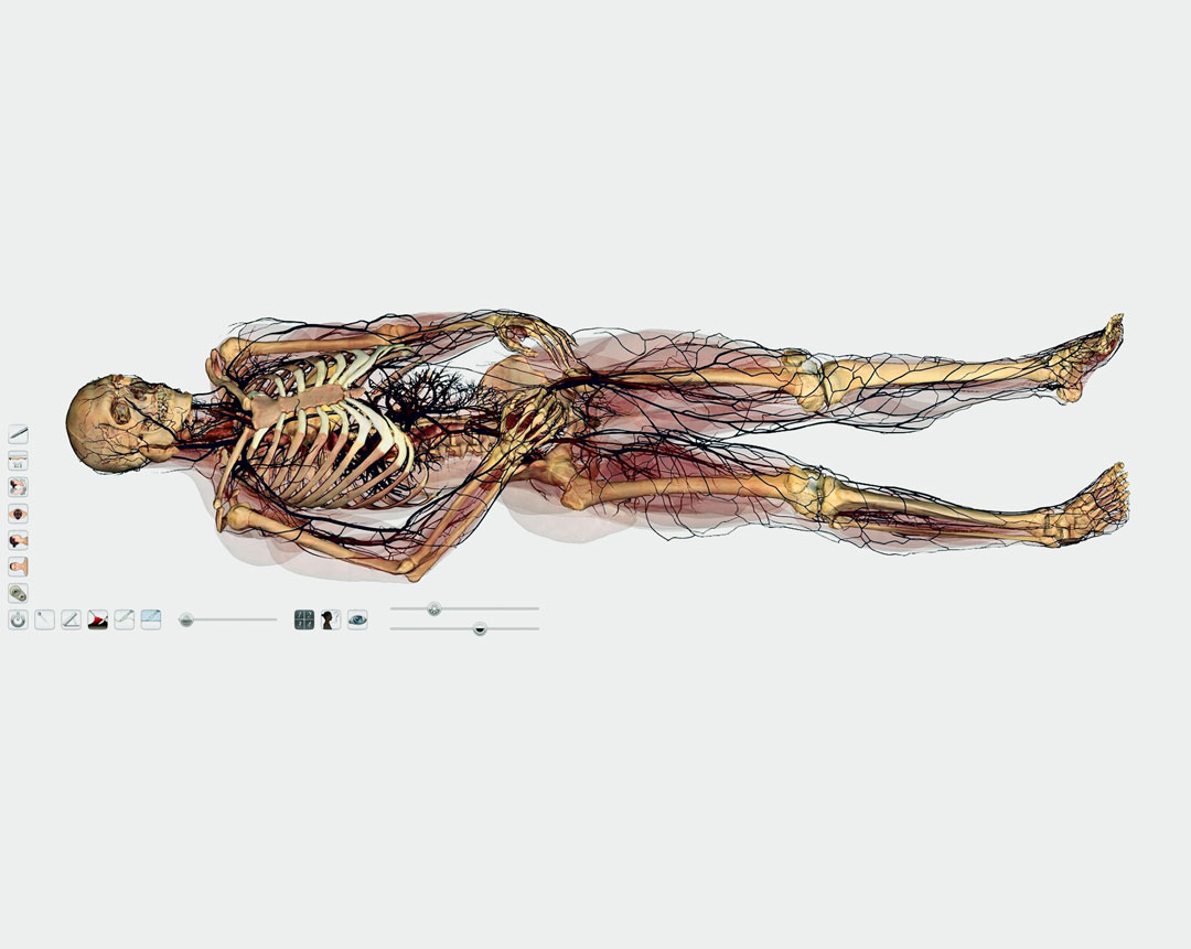
Today, they can explore and manipulate highly accurate three-dimensional scans, almost anywhere, thanks to products such as The Anatomage Table, the world’s first virtual dissection table.
“The Anatomage Table uses modern technology to visualize the human body on a large, flat, horizontal screen in such detail that it is sometimes referred to as ‘virtual dissection’,” explains our new book. “Computer imaging allows individual structures to be reconstructed in realistic three dimensions, so that each of the body systems can either be isolated or viewed in anatomical relation to each other.”
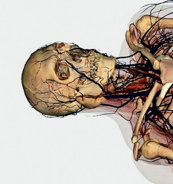
"Of course, while its pretty accurate, not everything on the table’s screen is exactly as you’d find it, were you to slice open a healthy human. “The body is coloured to mimic a traditional formalin-embalmed cadaver, with yellowy-pink bones and brown-purple muscles,” according to our new book. “The large muscle masses of the thighs and calves are well developed, but not well defined in detail. The cartilages of the ribcage stand out in stark white contrast to the uniform hues of the rest of the cadaver and the detail of the sternum reminds us that this is a reconstructed image. The impressive arborescent pattern of the blood vessels looks like a spider’s web spreading throughout every part of the body. The facial vessels are particularly striking as they run in an almost straight line, like a gash, from the inner corner of the eye down to the angle of the jaw.”
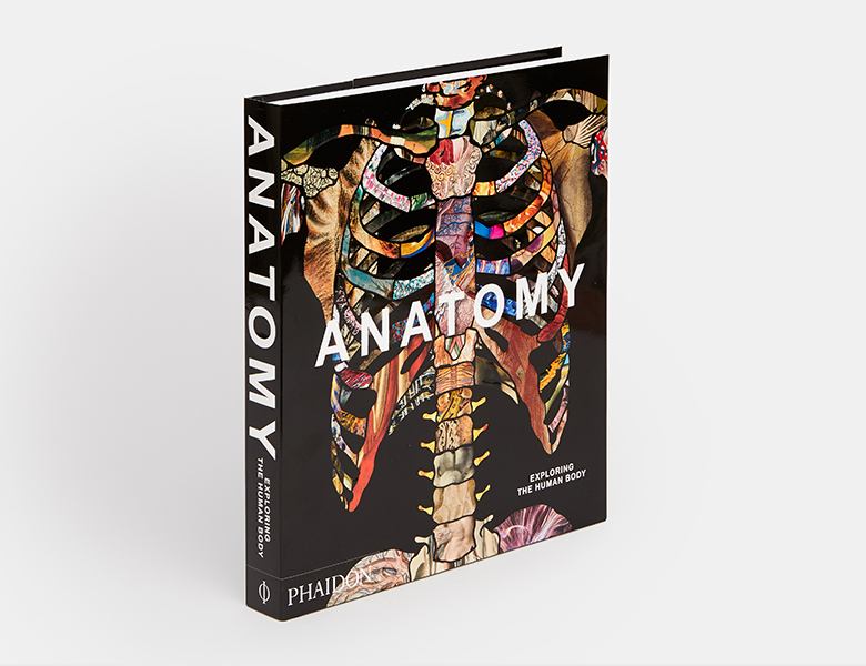
To see this image and many more that visualize the human body through the ages, get a copy of Anatomy here. The book is a visually compelling survey of more than 5,000 years of image-making. Through 300 remarkable works, selected and curated by an international panel of anatomists, curators, academics, and specialists, it chronicles the intriguing visual history of human anatomy, showcasing its amazing complexity and our ongoing fascination with the systems and functions of our bodies.
Exploring individual parts of the human body from head to toe, and revealing the intricate functions of body systems, such as the nerves, muscles, organs, digestive system, brain, and senses, this authoritative book presents iconic examples alongside rarely seen, breathtaking works. The 300 entries are arranged with juxtapositions of contrasting and complementary illustrations to allow for thought-provoking, lively, and stimulating reading. Order your copy here.