The Art of Anatomy – Eye Teaching Model
This 19th century model used a glass lens to demonstrate to both opticians and keen amateurs how our vision works
The London optician William Jones was not simply a maker of spectacles. He and his younger brother, Samuel, were among the city’s finest producers of scientific instruments during the late 18th and early 19th century, and exported their wares around the world. US Founding Father Thomas Jefferson bought a portable orrery – a miniature, mechanical model of the Solar System – from W. and S. Jones, and the company also supplied optical instruments to Harvard University.
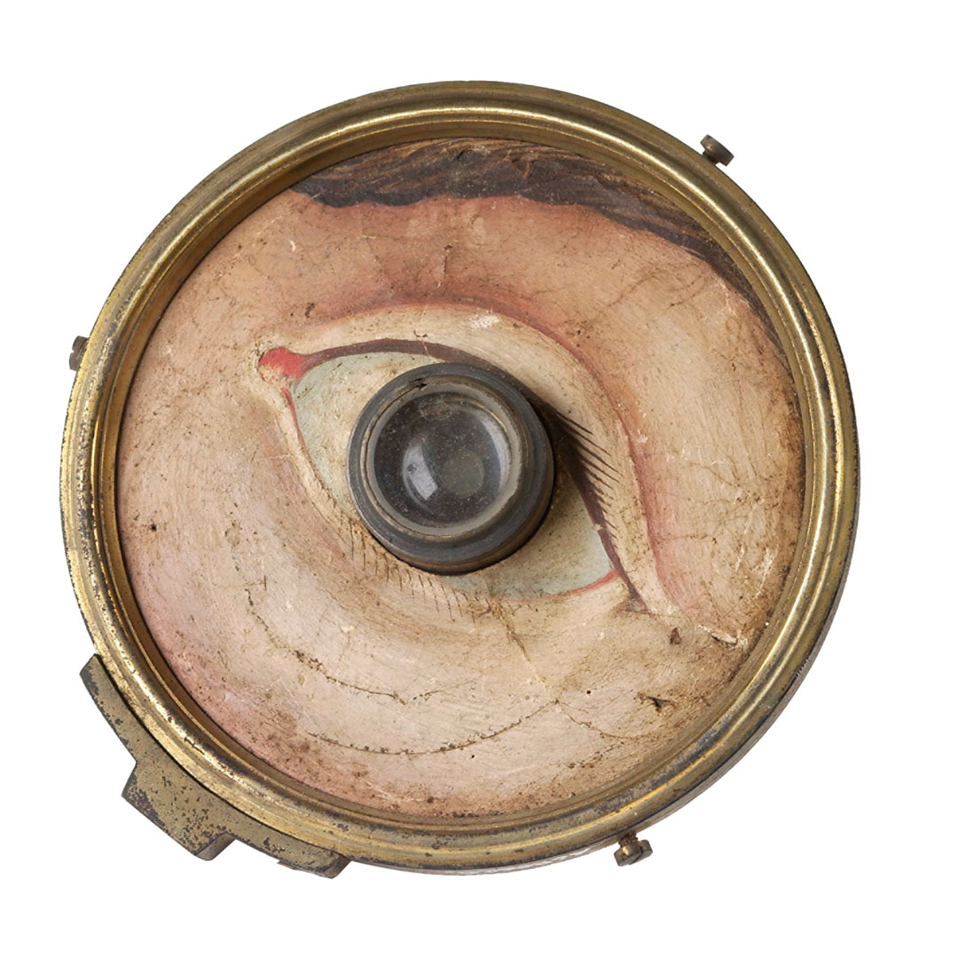
This particular creation – a small glass lens set into a brass backing, decorated with a paper, hand-painted eye socket – was also an educational instrument, though it may have been sold to consumers interested in how our vision works as a kind of philosophical toy too, explains our new book Anatomy: Exploring the Human Body.
“The optician or oculist who owned this model would shine a narrow beam of light – perhaps created with a candle – through a pinhole onto the glass ‘cornea’, and observe how the beam refracted, or bent, to reach the light-sensitive area known as the retina at the back of the eye,” explains our new book. “The model’s curved ‘retina’ is on the reverse side. A normal eyeball refracts light to a point exactly on the retina.
In a long eyeball, light is refracted in front of the retina, causing short-sightedness. Conversely, a long-sighted eyeball is short and refracts light to a theoretical point behind the retina. Practice eyes were sold with interchangeable corneas as well as corneal caps to represent spectacle lenses.” With a few, simple components, common malfunctions in human sight are made crystal clear.
This image is one of 300 remarkable works, selected and curated by an international panel of anatomists, curators, academics, and specialists in our new book Anatomy. The book is a visually compelling survey of more than 5,000 years of image making and chronicles the intriguing visual history of human anatomy, showcasing its amazing complexity and our fascination with the systems and functions of our bodies. The 300 entries are arranged with juxtapositions of contrasting and complementary illustrations that make for thought-provoking and lively reading. Buy your copy here.