Astonishing Animals – The Pigeon
Scott Echols's ghoulish bird image is the result of an innovative technology that helps us see inside animals
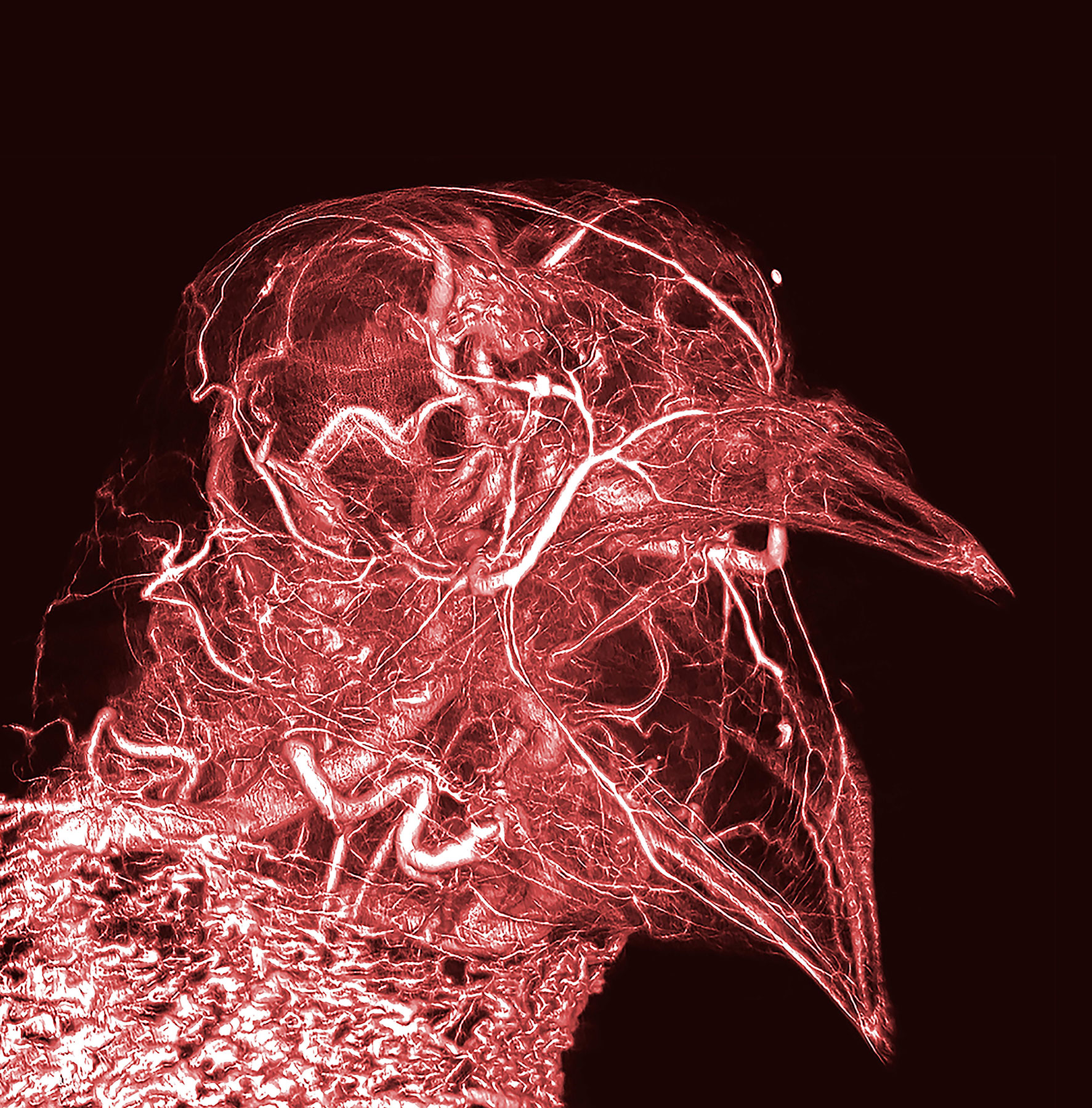
Despite its rather monstrous appearance, this image of a pigeon marks a significant progression for zoologists in visualising the internal anatomy of animals.
Having previously relied on sketching dissections or layering two-dimensional tissue structures together, this technically demanding image was created by flooding the animal’s arteries and veins with a contrast agent visible to X-rays.
This three-dimensional X-ray unveils the labyrinthine structure of veins, arteries and minuscule capillaries that carry blood through the head and shoulders of a common pigeon. The large arteries leading to the brain are clearly visible, as is the mass of veins in the pigeon’s neck that acts as a heart exchange when it engorges, allowing the animal to cool down via upper oesophageal pulsation – the bird equivalent of a dog’s panting.
The look of internal anatomy can be attained by other, more tangible means, such as the ‘plastination’ technique developed by German anatomy artist Gunther von Hagens, wherein the water and fat of bodily tissues are replaced by plastics. However, ‘digital dissections’ such as Echols’s pigeon have the upper hand: they don't have to be visited to be studied; just downloaded.
See more of the 300 plus ways we have documented the animals around us throughout time by ordering a copy of Animal: Exploring the Zoological World here. Check out our previous stories from the book on Sir Edwin Landseer's Monarch of the Glenn, Underwater photographer Alexander Semenov's Lion's Mane Jellyfish, Cai Guo-Qing's Heritage, Jill Greenberg's Diana Monkey Nick Veasey's Fruit Bat, The Sweat Bea and The Steppe Bison.